IMAGING-PAM
Version:
Chlorophyll Fluorescence Imaging System


IMAGING-PAM M-Series
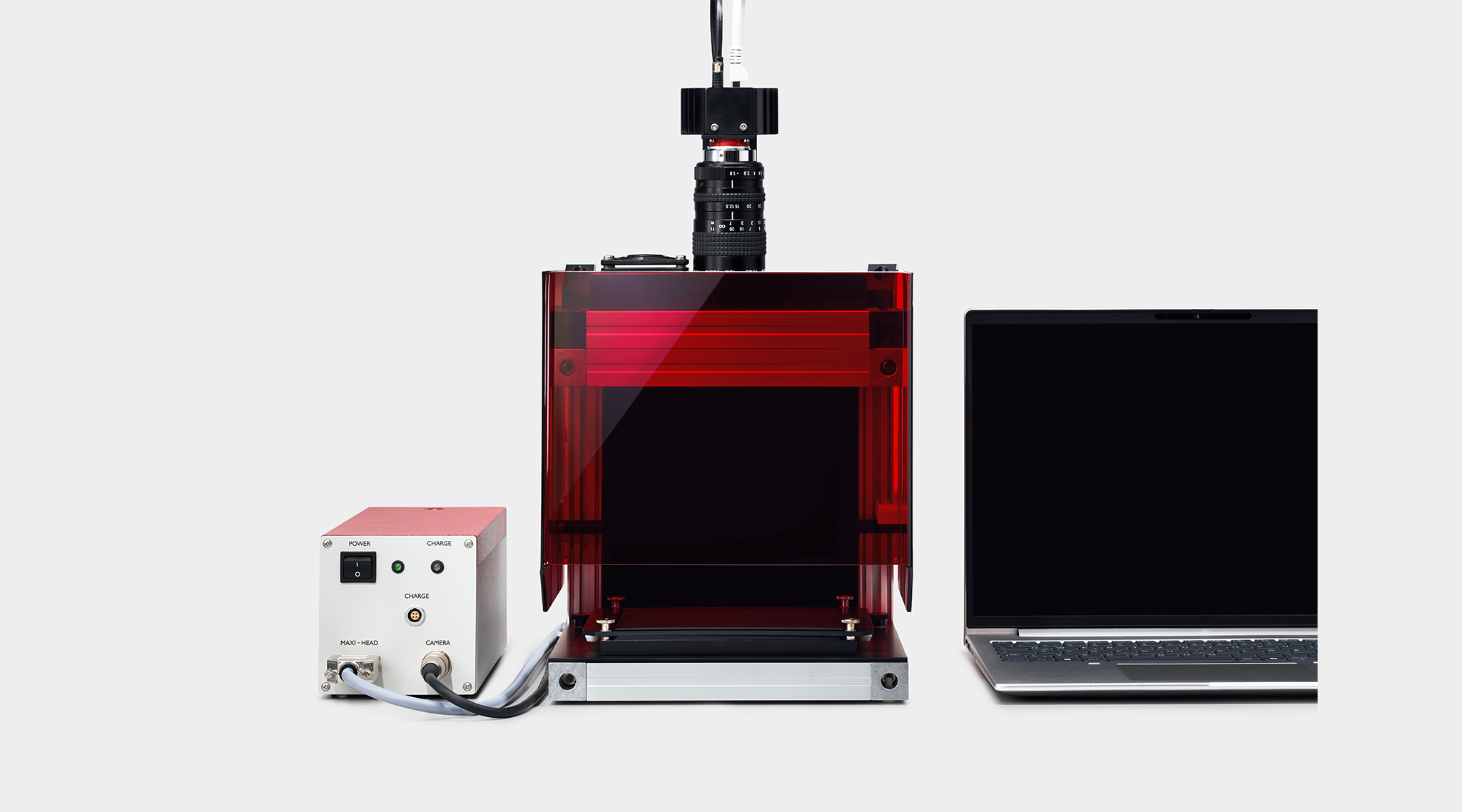
The IMAGING-PAM technology brings the power of the proven Pulse-Amplitude Modulation (PAM) principle into the realm of high-resolution, image-based chlorophyll fluorescence analysis. This approach delivers detailed, spatially precise insights into photosynthetic performance - non-invasive, highly sensitive, and ideal for capturing the dynamic responses of plants under real-world conditions. From fundamental plant physiology to advanced research on photosynthetic efficiency, stress resilience, and environmental toxicology, IMAGING-PAM systems offer researchers an exceptional window into the function and health of photosynthetic organisms.
The IMAGING-PAM M-series stands out through its modular, forward-looking design. All systems operate with the same Multi Control Unit IMAG-CG, and the camera can be used interchangeably across versions. This versatility enables seamless adaptation to different experimental requirements, applications, and magnifications, making the IMAGING-PAM M-Series both cost-effective and exceptionally flexible. In addition, all systems can be fully programmed for automated measurement routines and operated remotely via the ImagingWinGigE software, supporting long-term experiments and high-throughput workflows with minimal manual intervention.
What is Chlorophyll Fluorescence Imaging?
Conventional chlorophyll fluorescence measurements analyze a single point on a leaf, assuming the sample responds uniformly. In reality, stress, disease, and genotype differences rarely affect a leaf evenly. Chlorophyll fluorescence imaging overcomes this limitation by capturing the fluorescence signal across an entire sample area at once - visualizing the spatial distribution of photosynthetic performance pixel by pixel, rather than averaging it into a single number.
The IMAGING-PAM applies the same pulse-amplitude modulation (PAM) technique used in point-measuring fluorometers, but combines it with a sensitive camera that records the modulated fluorescence signal simultaneously across the entire imaging area. A defined sequence of measuring light, actinic light, and saturation pulses is applied to the whole sample, and the camera captures the resulting fluorescence at each pixel. This makes it possible to detect localized stress responses, early signs of pathogen infection, or genotype-specific differences long before they would be visible to the eye.
From this pixel-by-pixel data, the system automatically calculates false-color images of parameters such as FV/FM (maximum photochemical yield), Y(II) (effective photochemical yield), and NPQ (non-photochemical quenching) - turning complex physiological data into an intuitive, visual map of plant health across the entire sample.
Different versions for various applications
Choose your IMAGING-PAM. Select the version that fits your sample size, measurement context, and throughput requirements.
MAXI Version
Whole leaves, fruits, corals
The largest imaging area in the M-Series (10 × 13 cm²) with homogeneous LED illumination across the full measuring area. Can be combined with the Gas-Exchange Chamber 3010-GWK1 for simultaneous gas exchange.
MINI Version
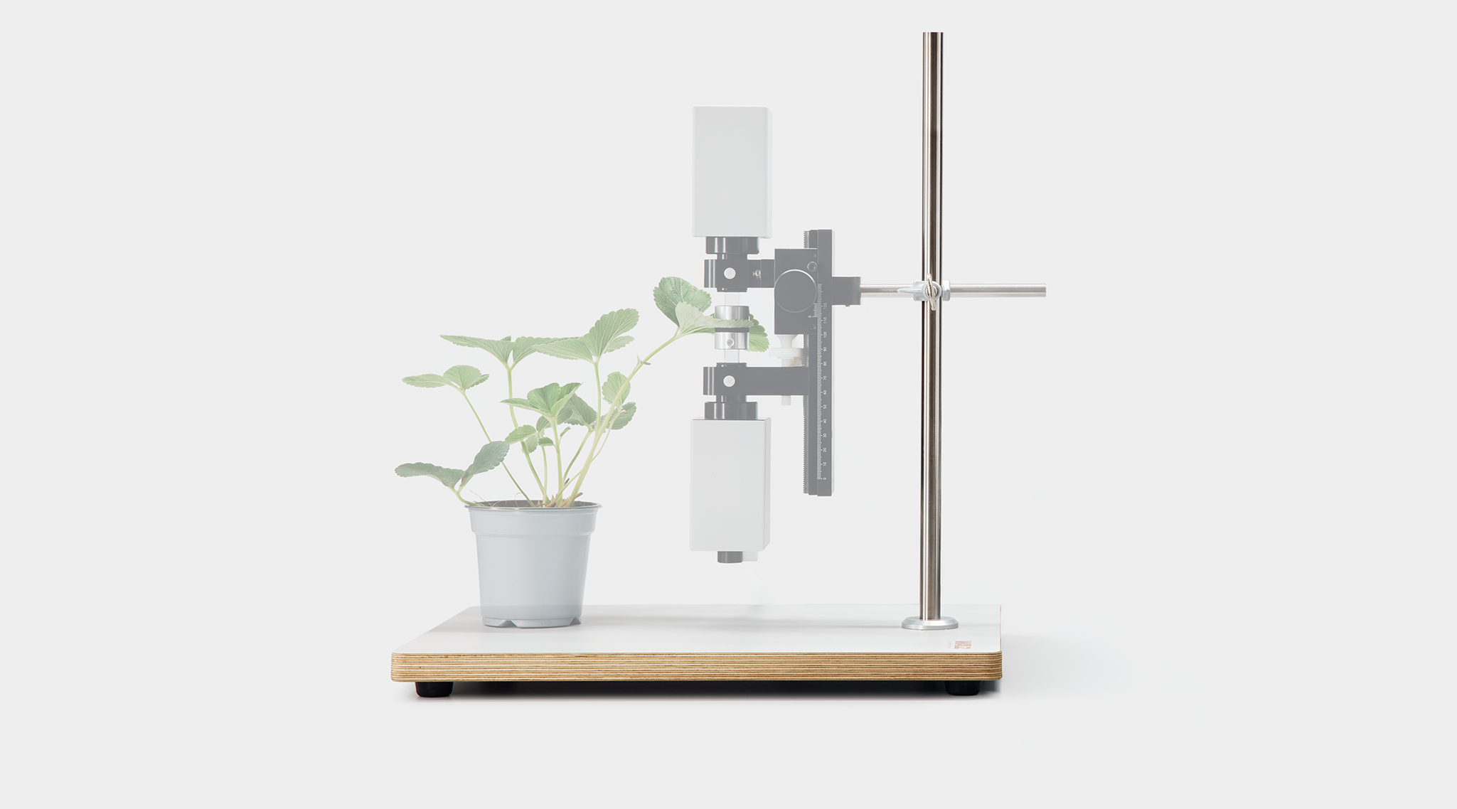
Field & single-leaf measurements
Compact and field-portable with a leaf clip holder for single-leaf measurements. Connects to the GFS-3000 via the snap-on Adapter IMAG-MIN/GFS for combined fluorescence imaging and gas exchange.
MICROSCOPY Version
Single cells, tissue sections
Integrates with a Zeiss Axioscope 5 microscope for cellular-level chlorophyll fluorescence imaging. The highest spatial resolution in the M-Series — ideal for microalgae, tissue sections, and single-cell analysis.
MOBILE Version
Field screening, flat surfaces
A lightweight, standalone imaging head for chlorophyll fluorescence measurements on flat surfaces. Designed for rapid screening in the field or greenhouse without requiring a full laboratory setup.
3D Version
Rosettes, shoots, canopies
Captures chlorophyll fluorescence images on non-flat, three-dimensional plant surfaces. Extends imaging capability beyond planar samples to whole shoots, rosettes, and complex canopy structures in the field.
HEXAGON-IMAGING-PAM
High-throughput screening
For automated, multi-sample screening applications with a large-area imaging platform (20 × 24 cm). Designed for plant phenotyping and high-throughput workflows available as a separate standalone instrument.
General Features IMAGING-PAM M-Series
Excitation Wavelengths and Filter Sets
The MAXI, MINI, and MICROSCOPY versions are available with blue, red-orange, and GFP-specific LEDs and dedicated filter sets. Blue excitation is standard for higher plants and algae, red-orange is recommended for cyanobacteria.
Actinic Light Intensity
High-performance power LEDs deliver up to 5,000 µmol m⁻² s⁻¹ (depending on version and configuration) for precise simulation of high-light stress and dynamic illumination conditions.
Ambient Light Compatibility
The systems support measurements under continuous ambient light. IMAGING-PAM devices communicate directly with the Universal Light Meter ULM-500 to automatically integrate real-time ambient PAR values into the report file.
Suspensions and Combined Techniques
Additional filter plates improve image quality for liquid suspension measurements through reflective surfaces. Dedicated adapters enable combined PSII imaging and gas-exchange analysis under controlled environmental conditions.

Application for IMAGING-PAM
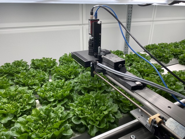
Development of an AI-based, energy-optimized illumination system for urban indoor plant cultivation
WALZ in collaboration with the Federal Agency for Agriculture and Food (BLE), INUGA, the Fraunhofer UMSICHT, University of Applied Sciences Osnabrück and Computomics
The challenges faced by agricultural systems are increasing globally. Current studies indicate that reducing negative environmental impacts while maintaining yield and striving for higher product quality pose significant challenges. The central goal of LightSaverAI is to establish the foundation for an intelligent production system for indoor farming in urban spaces. This system measures chlorophyll fluorescence (ChlFl) as an indicator of photosynthetic rate, along with various environmental parameters, and analyzes them using AI approaches. As a result, the real-time light requirements of plants are assessed, and an LED illumination module is adjusted through a feedback loop to provide continuous illumination tailored to growth phases and environmental conditions. Leveraging LED technology, this system achieves maximum photosynthetic rates with minimal energy consumption.
Learn more about: INUGA: LightSaverAI
Key features of the system
- Customized, resource-efficient plant illumination
- Capture and AI-based analysis of chlorophyll fluorescence and environmental parameters
- Expected outcomes and applications:
Software for improved plant breeding and monitoring, leading to resource savings in indoor farming
Enhanced use of image and data processing in horticulture
Transferability to other production sectors (vegetable cultivation, spice and tea production, pharmaceutical industry)
Project participants:
- Fraunhofer UMSICHT
- Hochschule Osnabrück (Osnabrück University of Applied Sciences)
- Computomics GmbH
- Heinz Walz GmbH (Associate)
Project details:
- Coordinator: Dr.-Ing. Dennis Schlehuber
- Duration: April 15, 2022, to April 14, 2025
WALZ is proud to introduce the Phenoplate, an innovative tool designed to uncover the intricate relationship between photosynthesis, light, and temperature in real time. With its seamless integration of the MAXI-IMAGING-PAM and a thermocycler, the Phenoplate enables high-throughput measurements and dynamic control of temperature, providing invaluable insights into how various organisms respond to fluctuating environmental conditions. For a deeper look into this innovative approach, please refer to our technical documentation.
Phenoplate Key Features:
- Precision Control of Temperature and Light: Simultaneously measure Photosystem II efficiency (Y(II)) and non-photochemical quenching (NPQ) while controlling temperature changes rapidly, enabling studies across multiple temperature gradients.
- High-Throughput Capability: The Phenoplate’s ability to handle 96-well plates allows for the simultaneous assessment of numerous replicates, ensuring robust, reproducible data. Whether you're studying microalgae, corals, or terrestrial plants, the Phenoplate’s flexibility makes it an invaluable tool for diverse biological research.
- Applications in Multiple Research Areas: From studying coral bleaching stress responses to examining how temperature and chemical gradients affect photosynthetic processes in microalgae, the Phenoplate provides a unique platform to capture key data for understanding photosynthesis in dynamic environments.

A Game Changer for Photosynthesis Research:
The Phenoplate isn’t just an instrument; it’s a powerful research tool designed to unlock the full potential of your experiments. By combining controlled environments with high-throughput capacity, the Phenoplate offers a precise and solution for researchers studying the effects of environmental stress on photosynthetic organisms. The innovation was developed by scientists from the Climate Change Cluster at University of Technology Sydney.


Scientific Publications using Walz Devices
Source: Google Scholar.
Keywords: (Walz OR Waltz) Effeltrich.
Date: June 22, 2026.
Ʃ = 19642
Source: Google Scholar.
Keywords: (Walz OR Waltz) Effeltrich.
Date: June 22, 2026.
Ʃ = 19642
Selected Publications
PFAS-herbicide diflufenican reduces the photosynthetic capacity in seagrass (Zostera marina L.)
Marine Environmental Research 210: 107342 [MINI]
UV RESISTANCE LOCUS 8 signalling enhances photosynthetic resilience to herbicide-induced damage in Arabidopsis thaliana
New Phytologist 247: 1763-1776 [MAXI]
Photosynthetic capacity and pigment distribution of a siphonous green alga, Dichotomosiphon tuberosus
Photosynthesis Research 163: 30 [MAXI]
Bioprinted photosynthetic living materials
PhD Thesis University of California San Diego [MINI]
The chloroplast RNA-binding protein CP29A supports rbcL expression during cold acclimation
Proceedings of the National Academy of Sciences USA 122: e2403969122 [MAXI]
Monitoring of spatial heterogeneity of chlorophyll fluorescence of cotton leaves at the early stage of Verticillium wilt based on spectral imaging
Industrial Crops & Products 226: 120663 [MAXI]
Chlorophyll fluorescence responses to CO2 availability reveal crassulacean acid metabolism in epiphytic orchids
Journal of Plant Research 138: 323-326 [MAXI]
Resistance to the herbicide metribuzin conferred to Arabidopsis thaliana by targeted base editing of the chloroplast genome
Plant Biotechnology Journal 23: 204-215 [MAXI]
The vacuolar inosital transporter BvINT1;1 contributes to raffinose biosynthesis and reactive oxygen species scavenging during cold stress in sugar beet
Plant, Cell & Environment 48: 3471-3486 [MINI]
Influences of environmental and leaf functional traits variations on photosynthetic characteristics of Cotoneaster multiflorus in Xinglong Mountain
Frontiers in Plant Science 16: 1562491 [MAXI]
Photoprotective strategies in pale versus melanic boreal hair lichens: non-photochemical quenching compensates for less protective fungal pigments
Planta 262: 3 [MAXI]
Synchotron macro-ATR-FTIR: a powerful technique for analyzing changes in plant cell chemical composition after surfactant exposure
The Plant Journal 122: e70227 [MINI]
Correlation analysis of twig and leaf characteristics and leaf thermal dissipation of Hippophae rhamnoides in the riparian zone of the Taohe River in Gansu Province, China
Plants 14: 282 [MAXI]
Integrating transcriptomics and metabolomics to comprehensively analyze phytohormone regulatory mechanisms in Rhododendron chrysanthum Pall. under UV-B radiation
International Journal of Molecular Sciences 26: 1545 [MAXI]
A novel method for measuring heat injury in leaves provides insights into the sequence of processes of heat injury development
Plant Methods 21: 89 [MAXI]
Methyl jasmonate was involved in hydrogen sulfide-alleviated cadmium stress in cucumber plants through ROS homeostasis and chlorophyll metabolism
International Journal of Molecular Sciences 26: 475 [MAXI]
In situ cavitation bubble manometry reveals a lack of light-activated guard cell turgor modulation in bryophytes
Proceedings of the National Academy of Sciences USA 122: e2419887122 [MINI]
The extended photoperiod impacts on sweet Basil (Ocimum basilicum) in natural tropical greenhouse
Horticulturae 11: 324 [MAXI]
Postharvest dynamics of photosynthesis in fresh-cut lettuce
Physiologia Plantarum 177: e70433 [MAXI]
Differentiation trajectory of virus-induced tumour cells in rice revealed by single-cell RNA sequencing
Plant Biotechnology Journal 23: 4794-4812 [MINI]
Genome-wide identification of APX genes in flax (Linum usitatissimum) and functional characterization of LuAPX12 in osmotic and salinity stress responses
BMC Plant Biology 25: 939 [MAXI]
Multi-omics research reveals the effects of the ABA-regulated phenylpropanoid biosynthesis pathway on the UV-B response in Rhododendron chrysanthum Pall.
Plants 14: 101 [MAXI]
Response of cool-season turfgrass monocultures and two-way mixtures to sequential acute drought periods
Crop Science 65: e21385 [MAXI]
Temperature-dependent responses of the hard corals Acropora sp. and Pocillopora verrucosa to molecular hydrogen
PLoS ONE 20: e0308894 [MAXI]
Sowing methods and strigolactones alleviate damage to the photosynthetic system of rice seedlings under salt stress by enhancing antioxidant capacity
Antioxidants 14: 1020 [MINI]
Anthropogenic impacts on coral-algal interactions of the subtropical lagoonal reef, Norfolk Island
Integrative Organismal Biology 7: obaf004 [MAXI]
Synergistic effects of supplemental lighting and foliar phosphorus application on flowering in passion fruit (Passiflora edulis)
Horticulturae 11: 478 [MAXI]
Light response of karyostrophy in the benthic pennate diatom Pleurosigma strigosum (Bacillariophyceae): a complementary photoprotective process?
Journal of Phycology 61: 1140-1152 [MICROSCOPY]
Genome-wide identification and expression divergence of CBF family in Actinidia arguta and functional analysis of AaCBF4 under cold stress
Life 15: 227 [MAXI]
Phototropin connects blue light perception to starch metabolism in green algae
Nature Communications 16: 2524 [MAXI]
Roles of vertical light-conducting carriers applied in microalgal-bacterial biofilm for enhanced nitrogen and phosphorus removal
Bioresource Technology 441: 133633 [MAXI]
Effects of high light intensity and spectral variability on maize photosynthesis and growth
Frontiers in Plant Science 16: 1511768 [MAXI]
Ice and air: visualisation of freeze-thaw embolism and freezing spread in young L. tulipifera leaves.
Journal of Experimental Botany 76: 5573-5587 [MINI]
Chloroplast precursor protein preClpD overaccumulation triggers multilevel reprogramming of gene expression and a heat shock-like response
Nature Communications 16: 3777 [MAXI]
Arbuscular mycorrhizal fungi enhance the saline-alkali tolerance of apple rootstock M9-T337 by regulating chlorophyll fluorescence parameters and hormone balance
Fruit Research 5: e0104 [MAXI]
Recovery of the cortical chloroplast layer in the green alga Chara after local irradiation
Frontiers in Plant Science 16: 1544999 [MICROSCOPY]
Single-cell sequencing reveals dynamic cell development trajectories in two kiwifruit (Actinidia chinensis) genotypes with contrasting cold resistance
Horticulture Advances 3: 24 [MAXI]
Chloroplastic aspartyl-tRNA synthetase is requirede for chloroplast development, photosynthesis and photorespiratory metabolism
Plant, Cell & Environment 48: 2998-3011 [MAXI]
Heat-evolved microalgae (Symbiodiniceae) are stable symbionts and influence thermal tolerance of the sea anemone Exaiptasia diaphana
Environmental Microbiology 27: e70011 [MAXI]
A Mg-chetalase subunit I missense mutant in barley exhibits a cold sensitive phenotype under field conditions
Physiologia Plantarum 177: e70434 [MAXI]
Mitigating algal competition with fouling-prevention coatings for coral restoration and reef engineering
ACS Sustainable Chemistry & Engineering 13: 5808-5817 [MINI]
Functional relationship of atypical thioredoxins with NADPH-thioredoxin reductase C and 2-Cys periredoxins in Arabidopsis chloroplasts
Journal of Experimental Botany 76: 5481-5498 [MAXI]
Transcriptomic and metabolomic evidence reveal the vital role of lactose in the acquistion of rapid dessication tolerance in Boea hygrometrica
Plant, Cell & Environment 48: 4564-4584 [MAXI]
Assessment of the morphological features, physiological and photosynthetic activity of the different cell forms of Symbiodinaceae using microfluidic methods
Frontiers in Photobiology 3: 1645420 [MICROSCOPY]
Physiological and transcriptomic analysis of Spartina alterniflora in response to imazapyr acid stress
BMC Plant Biology 25: 630 [MAXI]
An antagonism between ethylene signaling and DNA methylation orchestrates the progression of leaf senescence in non-heading Chinese cabbage
Advanced Science 12: e14954 [MAXI]
Melatonin improves salt tolerance in tomato seedlings by enhancing photosystem II functionality and Calvin Cycle activity
Plants 14: 1785 [MAXI]
Protection of photosynthesis by UVR8 and Cryptochromes in Arabidopsis under blue and UV radiation
Plant, Cell & Environment 48: 6321-6335 [MAXI]
Loss of state transitions in Bryopsidales macroalgae and kleptoplastic sea slugs (Gastropoda, Sacoglossa)
Communications Biology 8: 869 [MINI]
Squalene acts as a feedback signaling molecule in facilitating bidirectional communication between tea plants
Science Advances 11: eads4888 [MAXI]
Optimizing growth, physiology, and saponin production in Primula veris L. through tailored LED light spectra for energy-efficient cultivation
Agronomy 15: 2184 [MAXI]
Autoendolithic coral symbionts are sheltered from UV radiation
Research Summit 2025 Universidade de Aveiro 107 [MICROSCOPY]
Guard-cell expression of abscisic acid resceptors for engineering water-use-efficient plants without trade-offs in growth
New Phytologist 248: 690-705 [MAXI]
Overexpression of StHsfA2 enhances thermotolerance and promotes tuberization in potato under high temperature through StSP6A
Plant Biotechnology Journal 23: 5045-5062 [MAXI]
Possible lessons of a model experiment: to what extent can UV activate the production of leaf phenolics in indoor plant cultivation?
Plant Physiology and Biochemistry 219: 109333 [MAXI]
Elevated temperature decreases stony coral tissue loss disease transmission, with little effect of nutrients
Scientific Reports 15: 22261 [MAXI]
Siphonous green macroalgae with contrasting capacities for the energy-dependent quenching, qE, rely on different photoprotective mechanisms
bioRxiv [MINI]
Effect of light intensity and light spectrum of LED light sources on photosynthesis and secondary metabolite synthesis in Ocimum basilicum
Plants 14: 1334 [MAXI]
Inhibiting inositol transport disrupts metabolite profiles and mimics heat stress in a model cnidarian-Symbiodiniaceae symbiosis
Communications Biology 8: 755 [MAXI]
Thermal stability changes of photosynthesis during osmotic and salt stress in wheat varieties cultivated in Central Europe and Mediterranean North Africa
Photosynthetica 63: 165-181 [MAXI]
Combined enhancement of ascorbic acid, beta-carotene and zeaxanthin in gene-edited lettuce
Plant Biotechnology Journal 23: 1954-1967 [MAXI]
PiERF1 regulates cold tolerance in Plumbago indica L. through ethylene signalling
Scientific Reports 15: 1735 [MAXI]
Integrative transcriptomic and metabolomic analysis elucidates the vital pathways underlying the differences in salt stress responses between two chickpea (Cicer arientinum) varieties
BMC Plant Biology 25: 903 [MAXI]
Additive effects of climate change-related stress factors in Fucus serratus and Fucus vesiculosus
Marine Ecology Progress Series 762: 13-26 [MAXI]
The compensatory response of photosystem II photochemistry to short-term insect herbivory is suppressed under water deficit
Insects 16: 984 [MINI]
Developmental and environmental effects on VTC2-dependent leaf ascorbate accumulation and functions
Journal of Experimental Botany 76: 3823-3833 [MAXI]
Jasmonates regulate light-induced patchoulol biosynthesis in Pogostemon cablin
Medicinal Plant Biology 4: e015 [MAXI]
The thylakoid lumen Deg1 protease affects non-photochemical quenching via the levels of violaxanthin de-epoxidase and PsbS
The Plant Journal 121: e17263 [MAXI]
StTCTP positively regulates StSN2 to enhance drought stress tolerance in potato by scavenging reactive oxygen species
International Journal of Molecular Sciences 26: 2796 [MAXI]
Rhizosphere and root-associated microbial community structures and plant physiology responses to large patch disease in zoysiagrass
Grass Research 5: e019 [MAXI]
Controlling lampenflora in heritage sites: in situ testing of polyoxometalate-ionic liquids in the Pommery Champagne cellar
ChemPlusChem 90: e202500043 [MAXI]
WRKY27-SPDS1 module of Ichang papeda (Citrus ichangensis) promotes cold tolerance by modulating spermidine content
Horticulture Research 12: uhaf065 [MAXI]
Photosynthetic activity in the heterotropic plant genus Cuscuta (Convolvulaceae) is modulated by phylogeny and ontogeny
Annals of Botany, in press [MINI/GFP]
Heat-tolerant algal symbiont may prevent extirpation of the threatenend elkhorn, Acropora palmata, in Florida during intensifying marine heatwaves
Coral Reefs 44: 953-965 [MAXI]
Recovery of five cool-season turfgrasses following long-term ice encasement
Crop Science 65: e70053 [MAXI]
Imaging PAM fluorometry reveals stable photosynthetic efficiency in multibiont symbiose on coral reefs
Frontiers in Marine Science 12: 1568287 [MAXI]
Selective nutrient incorporation may underestimate heterotrophy of a mixotrophic reef-building coral
Communications Biology 8: 1285 [MAXI]
Synergistic effects of Dysmorphococcus globus on selenium enrichment and astaxanthin accumulation
Foods 14: 3249 [MAXI]
Heterogeneity of photosynthetic light acclimation within single leaves of Fagus sylvatica
Trees 39: 104 [MAXI]
Light limitation and foliar pathogenic infection impact phloem anatomy and function in Pinus radiata D. Don
Plant, Cell & Environment 48: 6356-6372 [MAXI]
Transcriptomic analysis reveals the participation of NTRC in iron homeostasis in Arabidopsis
Physiologia Plantarum 177: e70203 [MAXI]
The photosynthetic response of the freshwater red alga Thorae okadae to environmental gradients of temperature, irradiance, dessication, and salinity: adaptations to its stream habitat
Journal of Applied Phycology 37: 2753-2768 [MINI]
Quantifying coral-algal interactions in an acidified ocean: Sargassum spp. exposure mitigates low pH effects on Acropora cervicornis health
Frontiers in Marine Science 12: 1487102 [MAXI]
Salt gradient-driven adaptation in okra: uncovering mechanisms of tolerance and growth regulation
Frontiers in Plant Science 16: 1648092 [MAXI]
AvERF73 positively regulates waterlogging tolerance in kiwifruit by participating in hypoxia response and mevalonate pathway
Horticultural Plant Journal 11: 162-174 [MAXI]
SICV affects starch metabolism by regulating SIBAM3 stability under low night temperature stress in tomatoes
Horticulture Research 12: uhaf233 [MAXI]
The ABF4-bHLH28-COMT5 module regulates melatonin synthesis and root development for drought tolerance in citrus
The Plant Journal 121: e70078 [MAXI]
RNA-seq analysis and candidate gene mining of Gossypium hirsutum stressed by Verticillium dahliae cultured at different temperatures.
Plants 13: 2688
Acclimation of intertidal macroalgae Ulva prolifera to UVB radiation: the important role of alternative oxidase.
BMC Plant Biology 24: 143
Integrated analysis of the physiological, transcriptomic and metabolomic responses of Neopophyra haitanensis after exposure to UV-B radiation: an energy metabolism perspective
Frontiers in Marine Science 11: 1372252
Tolerance enhancement of Dendrobium officinale by salicylic acid family-related metabolic pathways under unfavorable temperature. (Maxi)
BMC Plant Biology 24: 770
Supplementary low far-red light promotes proliferation and photosynthetic capacity of blueberry in vitro plantlets.
International Journal of Molecular Sciences 25: 688
Eco-design marine infrastructure: enhancing biofilm colonization on 3D printing concrete. (Maxi)
Rencontres de l’Ingénierie Maritime 2024
Phenology and the response of photosynthesis to irradiance and temperature gradient in the herbal drug red alga, Chondria armata (Rhodomelaceae, Ceramiales) from Kagoshima, Japan. (Mini)
Journal of Applied Phycology 36: 2139-2152
STIC2 selectively binds ribosome-nascent chain complexes in the cotranslational sorting of Arabidopsis thylakoid proteins.
The EMBO Journal 43: 4699-4719
The effects of elevated temperatures on the reproductive biology of a mediterranean coral, Oculina patagonica. (Maxi)
Oceans 5: 758-769
Extremely heat tolerant photosymbiosis in a shallow marine benthic foraminifera. (Maxi)
Scientific Reports 6: 30930
Mitochondrial oxidative phosphorylation (mtOXPHOS) serves as a sentinel to gauge fluctuations under heat stress in Arabidopsis thaliana elucidated by comparative transcriptomics. (Maxi)
Plant Stress 14: 100613
Rational design of ROS scavenging and fluorescent gold nanoparticles to deliver siRNA to improve plant resistance to Pseudomonas syringae. (Mini)
Journal of Nanobiotechnology 22: 446
A pgr5 suppressor screen uncovers two distinct suppression mechanisms and links cytochrome b6f complex stability to PGR5. (Hexagon)
The Plant Cell 36: 4245-4266
Ecotoxicological effects of suspended sediments on marine microalgae using flow cytometry and pulse-amplitude modulation (PAM) fluorometry.
Marine Pollution Bulletin 208: 116968
Modulation of photosystem II function in celery via foliar-applied salicylic acid during gradual water deficit stress. (Maxi)
International Journal of Molecular Sciences 25: 6721
Photoprotective mechanisms in Elysia species hosting Acetabularia chloroplasts shed light on host-donor compatibility in photosynthetic sea slugs. (Mini)
Physiologia Plantarum 176: e14273
The photoprotective behavior of a motile benthic diatom as elucidated from the interplay between cell motility and physiological responses to a light microgradient using a novel experimental setup. (Mini)
Microbial Ecology 87: 40
Molecular mechanisms of resistance against PS II-inhibiting herbicides in Amaranthus retroflexus from the Czech Republic. (Microscopy)
Genes 15: 904
Ubiquitin-mediated degradation of SIPsbS regulates low night temperature tolerance in tomatoes. (Maxi)
Cell Reports 43: 114757
Establishment of a flow-through system for the macrophyte growth inhibition test (OECD 239) including photosynthetic activity measurement to determine early effects. (Maxi)
Environmental Toxicology and Chemistry 43: 2589-2600
Resilience of Xanthoria parietina under Mars-like conditions: photosynthesis and oxidative stress response. (Maxi)
Planta 259: 25
The microphytobenthos are abundant and mediate key carbon fluxes in tropical mangroves. (Maxi)
Estuaries and Coasts 47: 963-980
The Rhododendron chrysanthum Pall.s’ acetylation modification of Rubisco enzymes controls carbon cycling to withstand UV-B stress. (Maxi)
Biomolecules 14: 732
Broad-spectrum ubiquitin/ubiquitin-like deconjugation activity of the rhizobial effector NopD from Bradyrhizobium (sp. XS1150). (Maxi)
Communications Biology 7: 644
A quick and effective method for thermostability differentiation in cucumber (Cucumis sativus L.). (Maxi)
Physiologia Plantarum 176: e14215
Comparative phytotoxicity of metallic elements on duckweed Lemna gibba L. using growth- and chlorophyll fluorescence induction-based endpoints. (Maxi)
Plants 13: 215
CsABCG11.2 mediates theanine uptake to alleviate cadmium toxicity in tea plants (Camellia sinensis).
Horticulture Advances 2: 19
Loss of cold tolerance is conferred by absence of the WRKY34 promotor fragment during tomato evolution. (Maxi)
Nature Communications 15: 6667
Impact of rare earth elements in sediments on the growth and photosynthetic efficiency of the benthic Myriophyllum aquaticum. (Maxi)
Journal of Soils and Sediments 24: 3814-3823
Phosphorylation of 399S at CsHsp70 of Cymbidium sinense is essential to maintain chlorophyll stability. (Maxi)
Plant Physiology and Biochemistry 211: 108518
Tropical bloom-forming mesoalgae Cladophoropsis sp. and Laurencia sp. – responses to ammonium enrichment and a simulated heatwave. (Maxi)
Journal of Phycology 60: 554-573
Enhanced cold tolerance with increased soil moisture: thermal tolerance variability among alpine Ranunculus species and hybrids in Kosciuszko National Park, Australia. (Maxi)
Field Studies in Ecology 5: 1
Classification of tomato seedling chilling injury based on chlorophyll fluorescence imaging and DBO-BiLSTM. (Maxi)
Frontiers in Plant Science 15: 1409200
Photoperiod and temperature interactions drive the latitudinal distribution of Laminaria hyperborea (Laminariales, Phaeophyceae) under climate change.
Journal of Phycology 60: 1237-1255
Unique photosynthetic strategies employed by closely related Breviolum minutum strains under rapid short-term cumulative heat stress.
Journal of Experimental Botany 75: 4005-4023
Thermal tolerance traits of individual corals are widely distributed across the Great Barrier Reef. (Maxi)
Proceedings of the Royal Society B 291: 20240587
Assessment of concrete bioreceptivity and model organism performance for use in algal biofilm green façade systems. (Mini)
11. Jahrestagung des DAfStb 159-166
Exogenous calcium alleviates the photosynthetic inhibition and oxidative damage of the tea plant under cold stress. (Maxi)
Horticulturae 10: 666
Comparative transcriptome analysis of salt-tolerant and -sensitive soybean cultivars under salt stress. (Maxi)
International Journal of Molecular Sciences 25: 9918
In vitro culture of Campomanesia pubescens under different light qualities: a morphoanatomical and physiological characterization. (Maxi)
Contribuciones a las Ciencias Sociales 17: 1-26
Transcription factors ABF4 and ABR1 synergistically regulate amylase-mediated starch catabolism in drought tolerance.
Plant Physiology 191: 591-609
Poly(ADP-ribose)-binding protein RCD1 is a plant PARylation reader regulated by photoregulatory protein kinases.
Communications Biology 6: 429
A new biotechnology for in-planta gene editing and its application in promoting flavonoid biosynthesis in bamboo leaves.
Plant Methods 19: 20
Local adaptation through countergradient selection in northern populations of Skeletonema marinoi.
Evolutionary Applications 16: 311-320
Chemical mutagenesis and thermal selection of coral photosymbionts induce adaptation to heat stress with trait trade-offs.
Evolutionary Applications 16: 1549-1567
CGL160-mediated recruitment of the coupling factor CF1 is required for efficient thylakoid ATP synthase assembly, photosynthesis, and chloroplast development in Arabidopsis.
The Plant Cell 35: 488-509
Bismuth exposure affects morpho-physiological performances and the ionomic profile in garden cress (Lepidium sativum L.) plants.
Frontiers in Environmental Science 11: 1221573
Weak acids produced during anaerobic respiration suppress both photosynthesis and aerobic respiration.
Nature communications 14: 4207
Ascorbate-glutathione cycle involving in response of Bangia fuscopurpurea (Bangiales, Rhodophyta) to hyposalinity.
Frontiers in Marine Sciences 10: 1174472
The role of the epidermal physode layer in UV protection of Fucus species.
Journal of Photochemistry and Photobiology 15: 100174
Influence of nitrogen on grapevine susceptibility to downy mildew.
Plants 12: 263
Survivability of the lichen Xanthoria parietina in simulated Martian environmental conditions.
Scientific Reports 13: 4893
Morphological, antioxidase, and photosynthetic responses in Liaodong elata (Aralia elata) under treatments of light intensity.
Research Square
The change in metabolic activity of a large benthic foraminifera as a function of light supply.
Scientific Reports 13: 8240
Photosynthesis in the biomass model species Lemna minor displays plant-conserved and species-specific features.
Plants 12: 2442
Increased drought resistance in state transition mutants is linked to modified plastoquinone pool redox state.
bioRxiv
An ancient metabolite damage-repair system sustains photosynthesis in plants.
Nature Communications 14: 3023
Metrics of coral microfragment viability.
bioRxiv
Multiple paths of plant host toxicity are associated with the fungal toxin cercosporin.
Plant, Cell & Environment 46: 2542-2557
(Z)-3-Hexenol integrates drought and cold stress signaling by activating abscisic acid glucosylation in tea plants.
Plant Physiology 193: 1491-1507
Effects of heat stress during seed filling stage on Brassica napus seed oil accumulation and chlorophyll fluorescence characteristics.
Phyton – International Journal of Experimental Botany 92: 333-348
A novel and high-throughput approach to assess photosynthetic thermal tolerance of kelp using chlorophyll a fluorometry.
Journal of Phycology 59: 179-192
Transcriptome-wide identification and functional characterization of CIPK gene family members in Actinidia valvata under salt stress.
International Journal of Molecular Sciences 24: 805
Environmental gradients reveal stress hubs pre-dating plant terrestrialization.
Nature Plants 9: 1419-1438
Antibiotics reduce Pocillopora coral-associated bacteria diversity, decrease holobiont oxygen consumption and activate immune gene expression.
Molecular Ecology 32: 4677-4694
Measuring photonics in photosynthesis: combined micro-fourier image spectroscopy and pulse amplitude modulated chlorophyll fluorimetry at the micrometre-scale.
Biomimetics 7: 107
AtRsmD is required for chloroplast development and chloroplast function in Arabidopsis thaliana.
Frontiers in Plant Science 13: 860945
Colonisation of artificial structures by primary producers: competition and photosynthetic behaviour.
Biofouling 38: 493-506
Desiccation tolerance in bryophytes relates to elasticity but is independent of cell wall thickness and photosynthesis.
Physiologia Plantarum 174: e13661
Melanisation in boreal lichens is accompanied by variable changes in non-photochemical quenching.
Plants 11: 2726
Enzymes degraded under high light maintain proteostasis by transcriptional regulation in Arabidopsis
Proceedings of the National Academies of Sciences USA 119: e2121362119
Exogenous melatonin improved photosynthetic efficiency of photosystem II by reversible phosphorylation of thylakoid proteins in wheat under osmotic stress.
Frontiers in Plant Science 13: 966181
Clay 3D printing as a bio-design research tool: development of photosynthetic living building components.
Architectural Science Review 65: 185-195
Rubredoxin 1 is required for formation of the functional photosystem II core complex in Arabidopsis thaliana.
Frontiers in Plant Science 13: 824358
Foliar-applied manganese and phosphorus in deficient barley: linking absorption pathways and leaf nutrients status.
Physiologia Plantarum 174: e13761
Microbiome signatures in Acropora cervicornis are associated with genotypic resistance to elevated nutrients and heat stress.
Coral Reefs 41: 1389-1403
Phase-selective synthesis of anatase and rutile TiO2 nanocrystals and their impacts on grapevine leaves: accumulation of mineral nutrients and triggering the plants defense.
Nanomaterials 12: 483
Phenoplate: an innovative method for assessing interacting effects of temperature and light on non-photochemical quenching in microalgae under chemical stress.
New Biotechnology 66: 89-96
New protective coatings against lampenflora growing in the Pommery Champagne cellar.
International Biodeterioration & Biodegradation 173: 105459
Isoprene enhances leaf cytokinin metabolism and induces early senescence.
New Phytologist 234: 961-974
Circadian and diel regulation of photosynthesis in the bryophyte Marchantia polymorpha.
Plant, Cell & Physiology 45: 2381-2394
Assessment of temperature optimum signatures of corals at both latitudinal extremes of the Red Sea.
Conservation Physiology 10: coac002
Photosynthetic performance of symbiont‑bearing foraminifera Heterostegina depressa affected by sunscreens.
Scientific Reports 12: 2750
Plastid anionic lipids are essential for the development of both photosynthetic and non-photosynthetic organs in Arabidopsis thaliana.
International Journal of Molecular Sciences 22: 4860
Influence of magnetic field with Schumann resonance frequencies on photosynthetic light reactions in wheat and pea.
Cells 10: 149
Photobiological effects on ice algae of a rapid whole-fjord loss of snow cover during spring growth in Langerlussuaq, a West Greenland fjord.
Journal of Marine Science and Engineering 9: 814
Prevalence and photobiology of photosynthetic dinoflagellate endosymbionts in the nudibranch Berghia stephanieae.
Animals 11: 2200
Dissection of the mechanism of growth inhibition resulting from loss of the PII protein in the cyanobacterium Synechococcus elongatus PCC 7942.
Plant & Cell Physiology 62: 721–731
NTRC effects on non-photochemical quenching depends on PGR5.
Antioxidants 10: 900
Effects of photoperiod and light spectra on growth and pigment composition of the green macroalga Codium tomentosum.
Journal of Applied Phycology 33: 471-480
Features and applications of a field imaging chlorophyll fluorometer to measure stress in agricultural plants.
Precision Agriculture 22: 947–963
Assessment of various toxicity endpoints in duckweed (Lemna minor) at the physiological, biochemical, and molecular levels as a measure of diuron stress.
Biology 10: 684
Inactivation of cytosolic FUMARASE2 enhances growth and photosynthesis under simultaneous copper and iron deprivation in Arabidopsis.
The Plant Journal 106: 766–784
The endosymbiotic coral algae symbiodiniaceae are sensitive to a sensory pollutant: artificial light at night, ALAN.
Frontiers in Physiology 12: 695083
A high-throughput method for measuring critical thermal limits of leaves by chlorophyll imaging fluorescence.
Functional Plant Biology 48: 634-646
Features and applications of a field imaging chlorophyll fluorometer to measure stress in agricultural plants.
Precision Agriculture 22: 947-963
Morphological and cytological observations of corolla green spots reveal the presence of functional chloroplasts in Japanese gentian.
PLoS ONE 15: e0237173
VENOSA4, a human dNTPase SAMHD1 homolog, contributes to chloroplast development and abiotic stress tolerance.
Plant Physiology 182: 721-729
Chilli veinal mottle virus HCPro interacts with catalase to facilitate virus infection in Nicotiana tabacum.
Journal of Experimental Botany 71: 5656-5668
Rethinking the influence of chloroplast movements on non-photochemical quenching and photoprotection.
Plant Physiology 183: 1213-1223
Chemical genetics approach identifies abnormal inflorescence meristem 1 as a putative target of a novel sulfonamide that protects catalase2-deficient Arabidopsis against photorespiratory stress.
Cells 9: 2026
HEBE, a novel positive regulator of senescence in Solanum lycopersicum.
Scientific Reports 10: 11021
Functional trade-off of hydration strategies in old forest epiphytic cephalolichens.
Fungal Biology 124: 903-913
Bioimaging techniques reveal foliar phosphate uptake pathways and leaf phosphorus status.
Plant Physiology 183: 1472-1483
Acclimation of Arabidopsis thaliana to low temperature protects against damage of photosystem II caused by exposure to UV-B radiation at 9 °C.
Plant Physiology and Biochemistry 134: 73-80
Effects of natural solar UV-B radiation on three Arabidopsis accessions are strongly affected by seasonal weather conditions.
Plant Physiology and Biochemistry 134: 64-72
Fluctuating light experiments and semi-automated plant phenotyping enabled by self-built growth racks and simple upgrades to the IMAGING-PAM.
Plant Methods 15: 156
Noninvasive phenotyping of plant-pathogen interaction: consecutive in situ imaging of fluorescing Pseudomonas syringae, plant phenolic fluorescence, and chlorophyll fluorescence in Arabidopsis leaves.
Frontiers in Plant Science 10: 1239
Exogenous glycinebetaine reduces cadmium uptake and mitigates cadmium toxicity in two tobacco genotypes differing in cadmium tolerance.
International Journal of Molecular Sciences 20: 1612
Red sea corals under artificial light pollution at night (ALAN) undergo oxidative stress and photosynthetic impairment.
Global Change Biology 25: 4194-4207
Strigolactone-induced senescence of a bamboo leaf in the dark is alleviated by exogenous sugar.
Journal of Pesticide Science 43: 173-179
Salinity reduces 2,4-D efficacy in Echinochloa crusgalli by affecting redox balance, nutrient acquisition, and hormonal regulation.
Protoplasma 255: 785-802
Morpho-anatomical and physiological differences between sun and shade leaves in Abies alba Mill. (Pinaceae, Coniferales): a combined approach.
Plant, Cell & Environment 41: 1683-1697
A Cu/Zn superoxide dismutase gene from Saussurea involucrate Kar. & Kir., SiCSD, enhances drought, cold, and oxidative stress in transgenic tobacco.
Canadian Journal Plant Science 97: 816-826
SNOWY COTYLEDON 2 promotes chloroplast development and has a role in leaf variegation in both Lotus japonicus and Arabidopsis thaliana.
Molecular Plant 10: 721-734
Mutation in Mg-protoporphyrin IX monomethyl ester cyclase decreases photosynthesis capacity in rice.
PLoS ONE 12: e0171118
Direct impact of the sustained decline in the photosystem II efficieny upon plant productivity at different developmental stages.
Journal of Plant Physiology 212: 45-53
Quantifying the efficiency of photoprotection
Philosophical Transactions of the Royal Society B 372: 20160393
Is colonization of sea ice by diatom facilitated by increased surface roughness in growing ice crystals?
Polar Biology 40: 593-602
Expanding the Symbiodinium (Dinophyceae, Suessiales) toolkit through protoplast technology.
Journal of Eukaryotic Microbiology 64: 588-597
The diatom Phaeodactylum tricornutum adjusts nonphotochemical fluorescence quenching capacity in response to dynamic light via fine-tuned Lhcx and xanthophyll cycle pigment synthesis.
New Phytologist 214: 205-218
Proteomic insight into the response of Arabidopsis chloroplasts to darkness.
PLoS ONE 11: e0154235
Microspatial variability in community structure and photophysiology of calcified macroalgal microbiomes revealed by coupling of hyperspectral and high-resolution fluorescence imaging.
Scientific Reports 6: 22343
Multiple impacts of loss of plastidic phosphatidylglycerol biosynthesis on photosynthesis during seedling growth of Arabidopsis.
Frontiers in Plant Science 7: 336
Global transcriptome analyses provide evidence that chloroplast redox state contributes to intracellular as well as long-distance signaling in response to stress and acclimation in Arabidopsis.
Photosynthesis Research 128: 287-312
HYPERSENSITIVE TO HIGH LIGHT1 interacts with LOW QUANTUM YIELD OF PHOTOSYSTEM II1 and functions in protection of Photosystem II from photodamage in Arabidopsis.
The Plant Cell 26: 1213‐1229
Moderate warming increases PS II performance, antioxidant scavenging systems and biomass production in Stylosanthes capitata Vogel.
Environmental and Experimental Botany 102: 58–67
Cyanobacteria in the Australian northern savannah detect the difference between intermittent dry season and wet season rain.
Biodiversity and Conservation 23: 1827‐1844
Effects of temperature and irradiance on a benthic microalgal community: A combined two-dimensional oxygen and fluorescence imaging approach
Limnology and Oceanography 59: 1599–1611
Removal of snow cover inhibits spring growth of Arctic ice algae through physiological and behavioral effects
Polar Biology 37: 471-481
Functional characteristics of a fruticose type of lichen, Stereocaulon foliolosum Nyl. in response to light and water stress.
Acta Physiologiae Plantarum 35: 1605‐1615
Chlorophyll fluorescence imaging: a new method for rapid detection of herbicide resistance in Alopecurus myosuroides.
Weed Research 53: 399-406
A thioredoxin‐like/β‐propeller protein maintains the efficiency of light harvesting in Arabidopsis.
Proc Natl Acad Sci USA 110: E2733–E2740
Photobiology of sea ice algae during initial spring growth in Kangerlussuaq, West Greenland: insights from imaging variable chlorophyll fluorescence of ice cores.
Photosynthesis Research 112: 103-115
Glycine decarboxylase controls photosynthesis and plant growth.
FEBS Letters 586: 3692–3697
Light‐dependent maintenance of hydraulic function in mangrove branches: do xylary chloroplasts play a role in embolism repair?
New Phytologist 195: 40–46
Photosynthetic limitations and volatile and non‐volatile isoprenoids in the poikilochlorophyllous resurrection plant Xerophyta humilis during dehydration and rehydration
Plant, Cell & Environment 35: 2061–2074
Combined effects of elevated CO2 and natural climatic variation on leaf spot diseases of redbud and sweetgum trees.
Environmental Pollution 158: 108-114
Mesoscale variation in the photophysiology of the reef building coral Pocillopora damicornis along an environmental gradient.
Estuarine, Coastal and Shelf Science 83: 186-196
A rapid, non-invasive procedure for quantitative assessment of drought survival using chlorophyll fluorescence.
Plant Methods 4: 27
A fluorescence-based bioassay for aquatic macrophytes and its suitability for effect analysis of non-photosystem II inhibitors.
Environmental Science and Pollution Research 14: 377-383
Zooxanthellae harvested by ciliates associated with brown band syndrome of corals remain photosynthetically competent.
Applied and Environmental Microbiology 73: 1968-1975
Deficiency in phylloquinone (vitamin K1) methylation affects prenyl quinone distribution, photosystem I abundance, and anthocyanin accumulation in the Arabidopsis AtmenG mutant.
Journal of Biological Chemistry 281: 40461-40472
A method for quantitative analysis of spatially variable physiological processes across leaf surfaces.
Photosynthesis Research 90: 161-172
Coral photobiology studied with a new imaging pulse amplitude modulated fluorometer.
Journal of Phycology 41: 335-342
Niche and photosynthesis of Chlorophyll d-containing cyanobacteria.
Nature 433: 820
Spatial heterogeneity of photosynthesis and the effect of temperature-induced bleaching conditions in three species of corals.
Marine Biology 144: 633-640
Complex regulation of gene expression, photosynthesis and sugar levels by pathogen infection in tomato.
Physiologia Plantarum 122: 419-428
IMAGING-PAM
Minimum PC requirements: Win10 or 11 OS, Intel Core i5-10xx or higher CPU, NVDIA® graphics board (mandantory, min 2 GB or more dedicated memory), SSD, min 8GB RAM, internal Gigabit Ethernet (GigE), HDMI interface.
The 3D part of the software can only be used under License. Therefore, a USB dongle is necessary (included in the basic instrument).
The LED array is mounted on a printed circuit board in an aluminium housing with a central opening for the IMAG-K6 and IMAG-K7 cameras. A fan on the top side provides the necessary cooling. Cable connections to the IMAG-CG control unit and the external 300 W power supply exit the rear of the housing. The IMAG-MAX/F filter plate is also recommended for measurements on reflective surfaces.
Light sources for fluorescence excitation and actinic illumination:
44 royal-blue 3 W Cree LEDs (450 nm), equipped with individual collimating optics; standard excitation intensity 0.5 µmol m-2 s-1 PAR, modulation frequency 1-8 Hz; maximum actinic intensity 1900 µmol m-2 s-1 PAR; maximum saturation pulse intensity 4000 μmol quanta µmol m-2 s-1 PAR.
Light sources for absorbed PAR and live video options:
16 red LEDs (660 nm); 16 NIR LEDs (780 nm)
17.5 cm x 18.5 cm x 4.5 cm (L x W x H)
1.3 kg (incl. 1.5 m long cable)
Standard 18.5 cm for 10 cm x 13 cm image area
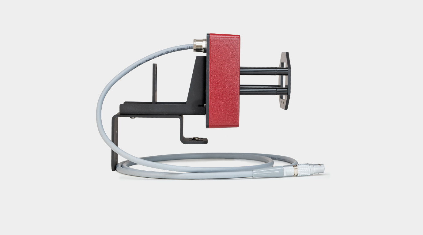
This mounting set is designed for the IMAG-K7 camera and can be used with the IMAG-MAX/L or IMAGMAX/LR. It consists of a camera holder and a 15 cm long metal rod (15 mm diameter) for mounting the LED array on a vertical stand. A double socket clamp and eye protection are required.
Aluminum frame with two clamps for secure mounting the LED-Array IMAG-MAX/L or IMAG-MAX/LR; red Perspex sliding hood for eye protection (a red version set will be shipped with a blue Perspex hood); removable bottom plate and four 20 cm housing legs are included.
Modified AxioScope 5 Microscope (Zeiss) adapted for IMAGING PAM applications. Comprises binocular fototube (30°/23 100:0/0:100), condenser 0,9/1,25H and transmitted light unit HAL 50. Detector filter RG665, dichroic mirror 420-640 nm, video adapter 60N-C 2/3" 0,5x and standard lens Fluar 20x are already mounted
For Windows 10 and 11 PCs with ImagingWinGigE software versions; connection is via GigE ethernet
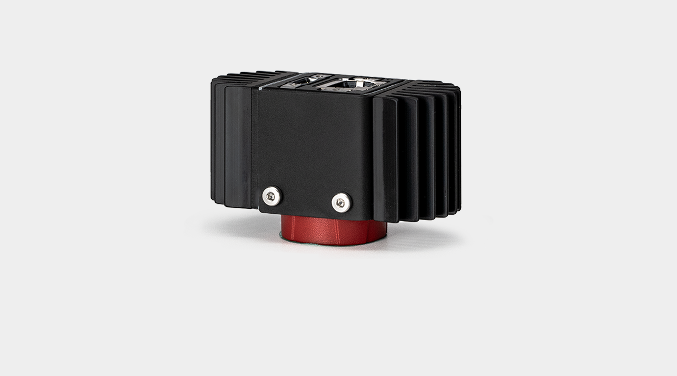
Four tilted LED arrays in a sturdy aluminum housing with a central opening for the IMAG-K7 or IMAG-K9 cameras. In a similar optical geometry, additional red and NIR LEDs are arranged around the central opening to ensure optimum illumination homogeneity. Anti-reflection filters matching the wavelength are already included and fitted.
Light sources for fluorescence excitation and actinic illumination:
12 Luxeon LEDs 460 nm with individual band pass filters and collimator optics; 10 red 650 nm and 10 NIR 780 nm LEDs for measuring PAR-absorptivity; max. actinic intensity 5000 μmol m-2 s-1 ; max. Saturation Pulse intensity, 10.000 μmol m-2 s-1 ; spacer frame at fixed working distance (7 cm); for imaging 24 mm x 32 mm sample area; suitable for use in combination with IMAG-K6 and IMAG-K7 with camera accessories (objectives and mounting set needed)
Black and white C-mount camera operated in 11-bit-mode at 16 frames/sec featuring 2 x 2 pixel binning
2/3" readout (1392 x 1040 px primary resolution; 640 x 480 px effective resolution after binning)
GigE-Vision®
C-mount
2.9 cm x 2.9 cm x 4.1 cm (L x W x H) (without lens and cooling bodies)
65 g (camera only)
Aluminum parts for mounting camera IMAG-K6 on the Imaging MINI-head LED arrays K6-MIN
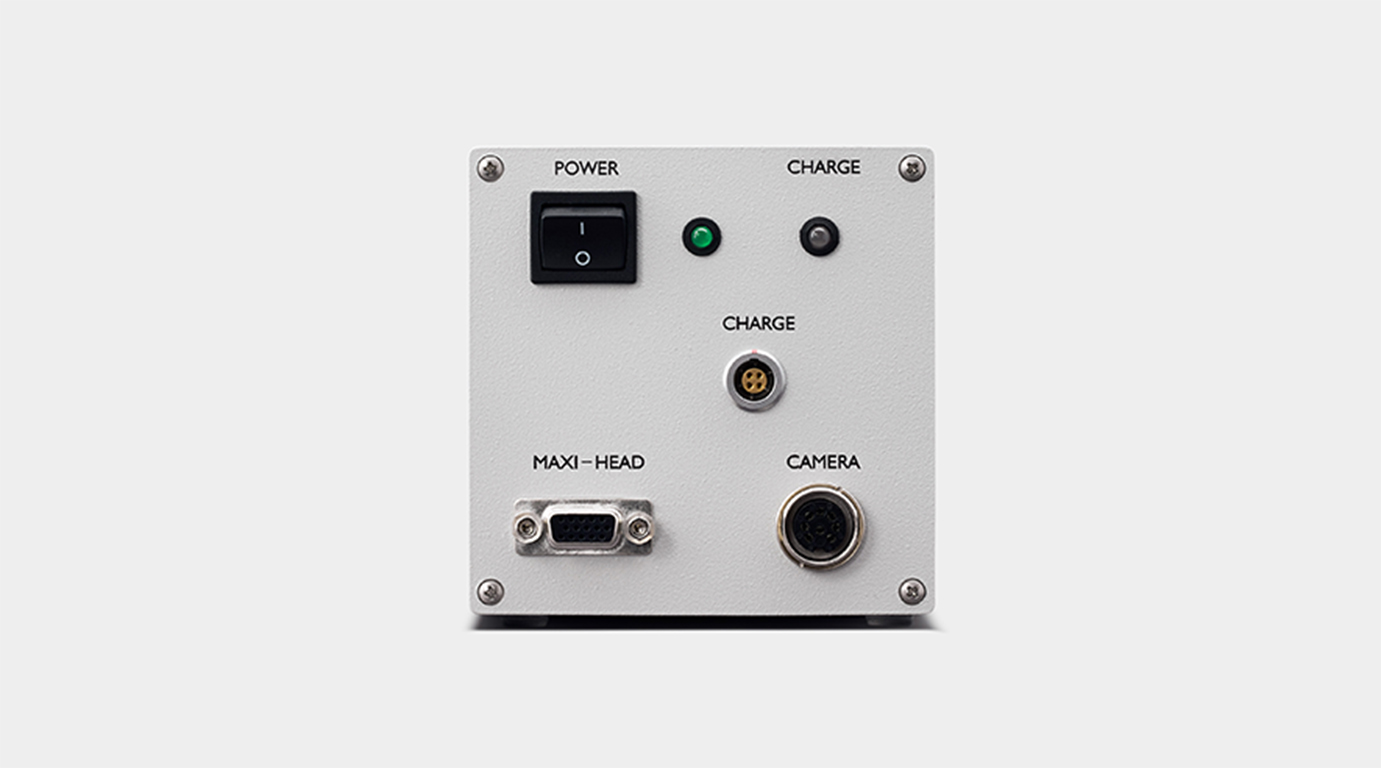
Aluminum housing featuring built-in Li-ion battery, sockets for cable connections with the cameras IMAG-K9, connectors for the MAXI or MINI Measuring Heads and trigger cable on one end for better portability and Battery Charger 2120-N
RISC processor
Light sources for fluorescence excitation and actinic illumination:
12 Luxeon LEDs 460 nm with individual band pass filters and collimator optics; 10 red 650 nm and 10 NIR 780 nm LEDs for measuring PAR-absorptivity; max. actinic intensity 5000 μmol m-2 s-1 ; max. Saturation Pulse intensity, 10.000 μmol m-2 s-1 ; spacer frame at fixed working distance (7 cm); for imaging 24 mm x 32 mm sample area; suitable for use in combination with IMAG-K6 and IMAG-K7 with camera accessories (objectives and mounting set needed)
Fully rugged tablet computer with sunlight-readable 11.6-inch sunlight-readable touchscreen including spare batteries and external charger. 8 GB RAM, 2.3 GHz processor or better, 10/100/1000 base Ethernet, GPS, Bluetooth (v4.2 class 1). OS Windows 11 64bit, ImagingWin GigE Software installed
Intel core i5 or comparable CPU, min 8 GB free RAM, built-in Gigabit Ethernet (GigE), Win11 OS
Adapter IMAG‐MAX/GWK1 for IMAGING-PAM (MAXI-Version) on Gas Exchange Chamber (3010-GWK1)
Adapter Plate with legs and eye protection for positioning IMAG-MAXI Head on 3010-GWK1
Mini quantum sensor for selective PAR (photosynthetically active radiation) measurement, cosine corrected for PPFD (photosynthetical photon flux density) measurement.
Black anodized aluminum housing
Perspex
High stability silicone photovoltaic detector with filter set for PAR correction (to learn more about the typical sensitivity see “General Features”). Signal output typically -2 μA / (1000 μmol m-2 s-1)
0.01 %/K
± 5 %
error < 4 % between angles from -80° to +80° from normal axis
Typically 1.32
- 5 °C … + 45 °C
3 m
BNC
Not required
Height: 16 mm
Diameter: 14 mm
Diffuser diameter: 5.5 mm
32 g
120 W AC adapter
PC-software ImagingWinGigE for Win 11
xpim, csv, jpg, tif (raw image format is b/w in 11-bit color depth, 1200 x 1000)
Intel core i5 or comparable CPU, min 8 GB free RAM, built-in Gigabit Ethernet (GigE), Win11 OS
Data display and evaluation plus instrument settings on 7 different windows
Blue Excitation Light Source: 451 nm dominant wavelength (ML, AL and SP) 6 x 13 Cree high power LEDs
Far Red light Source: 730 nm peak wavelength
0 – 50°C
C-mount
C-mount
Aluminum box with individual foam lining for IMAGING-PAMs and accessories
60 cm x 40 cm x 35 cm (L x W x H)
5 kg
Aluminum box with individual foam lining for IMAGING-PAMs and accessories
60 cm x 40 cm x 35 cm (L x W x H)
5 kg
Aluminum box with individual foam lining for IMAGING-PAMs and accessories
60 cm x 40 cm x 35 cm (L x W x H)
5 kg
Box1: Sturdy cardboard box with individual foam lining for IMAGING-3D and accessories
Box 2: Aluminum box with individual foam lining for IMAGING-PAM accessories
Box 1: 70 cm x 55 cm x 45 cm (L x W x H)
Box 2: 60 cm x 40 cm x 35 cm (L x W x H)
Box 1: 7 kg
Box 2: 5 kg
Aluminum box with individual foam lining for HEXAGON-IMAGING-PAM and accessories
62 cm x 62 cm x 62 cm (L x W x H)
6 kg
Box 1: Aluminum box with individual foam lining for IMAGING-PAM MICROSCOPY version accessories
Box 2: Sturdy cardboard box with individual foam lining for Zeiss Axioscope 5 and accessories
Box 1: 60 cm x 40 cm x 35 cm (L x W x H)
Box 2: 68 cm x 62 cm x 64 cm (L x W x H)
Box 1: 5 kg
Box 2: 5 kg
Accessories
This open leaf holder is used to fix sample leaves at a defined working distance of 18.5 cm, perpendicular to the optical axis - for mounting LED-Array Illumination Unit IMAG-MAX/L or IMAG-MAX/LR. It should only be used when additional eye protection is assured.
44 individual blue filters in black-anodized aluminum plate to be mounted in front of collimating optics of IMAG-MAX/L (not compatible with IMAG-MAX/LR). Recommended for the work with reflecting/wet surfaces. Not compatible with the red LED array illumination unit IMAG-MAX/LR.
The LED array is mounted on a printed circuit board in an aluminium housing with a central opening for the IMAG-K6 and IMAG-K7 cameras. A fan on the top side provides the necessary cooling. Cable connections to the IMAG-CG control unit and the external 300 W power supply exit the rear of the housing. An appropriate filter plate is included; not compatible with filter plate IMAG-MAX/F
Light sources for fluorescence excitation and actinic illumination:
44 red 3 W Luxeon LEDs (650 nm) equipped with individual collimating optics; standard excitation intensity 0.5 µmol m-2 s-1 PAR, modulation frequency 1-8 Hz; max. actinic intensity 1300 µmol m-2 s-1 PAR; max. saturation pulse intensity 3700 μmol µmol m-2 s-1 PAR.
Light sources for absorbed PAR and live video options:
16 red LEDs (660 nm); 16 NIR LEDs (780 nm)
Standard 18.5 cm for 10 cm x 13 cm image area
17.5 cm x 18.5 cm x 4.5 cm (L x W x H)
1.3 kg (incl. 1.5 m long cable)
MINI-Heads; MINI-Head holding grip
Four tilted LED arrays in a sturdy aluminum housing with a central opening for the IMAG-K6 camera. In a similar optical geometry, additional red and NIR LEDs are arranged around the central opening to ensure optimum illumination homogeneity. For GFP or PS II measurement the additional filter set K6-MIN/FS is necessary
12 Luxeon LEDs 480 nm with individual band pass filters and collimator optics for best combined excitation of GFP and PS II; 10 red 650 nm and 10 NIR 780 nm LEDs for measuring PAR-absorptivity; spacer frame at fixed working distance (7 cm); for imaging 24 mm x 32 mm sample area; suitable for use in combination with IMAG-K6 and IMAG-K7 with camera accessories (objectives and mounting set needed). Filter slider IMAG-MIN/FS necessary for GFP detection. Compatible with IMAG-K6 camera
Adapter IMAG‐MIN/GFS for IMAGING-PAM (MINI-Version) on Gas Exchange Standard Measuring Head (3010-S)
Four tilted LED Arrays in a sturdy aluminum housing with a central opening for the IMAG-K7 or IMAG-K9 cameras. In a similar optical geometry, additional red and NIR LEDs are arranged around the central opening to ensure optimum illumination homogeneity. Anti-reflection filters matching the wavelength are already included and fitted.
Light sources for fluorescence excitation and actinic illumination:
12 Luxeon LEDs 620 nm with individual short pass filters and collimator optics; 10 red 650 nm and 10 NIR 780 nm LEDs for measuring PAR-absorptivity; max. actinic intensity 2800 μmol m-2 s-1 ; max. Saturation Pulse intensity, 7700 μmol m-2 s-1 ; spacer frame at fixed working distance (7 cm); for imaging 24 mm x 32 mm sample area; suitable for use in combination with IMAG-K6 and IMAG-K7 with camera accessories (objectives and mounting set needed).
Red-Green-Blue Microscopy LED Lamp allowing computer-assisted deconvolution of major algae groups. Fluorescence excitation and actinic illumination using red (620 nm), green (520 nm), blue (460 nm) or white light (mixed 620, 520 and 460 nm); featuring fluid light guide (100 cm length, 3 mm active dia.), connecting to collimator optics at excitation port of epifluorescence microscope; with cable to be connected to RGB output socket at IMAG-CG; featuring printed circuit board with separate drivers for RGB LEDs;
F1.4/f=16 mm lens with detector filter (RG645, 3 mm) and short-pass interference filter
C-Mount
42 x 50 mm
155 g (lens only)
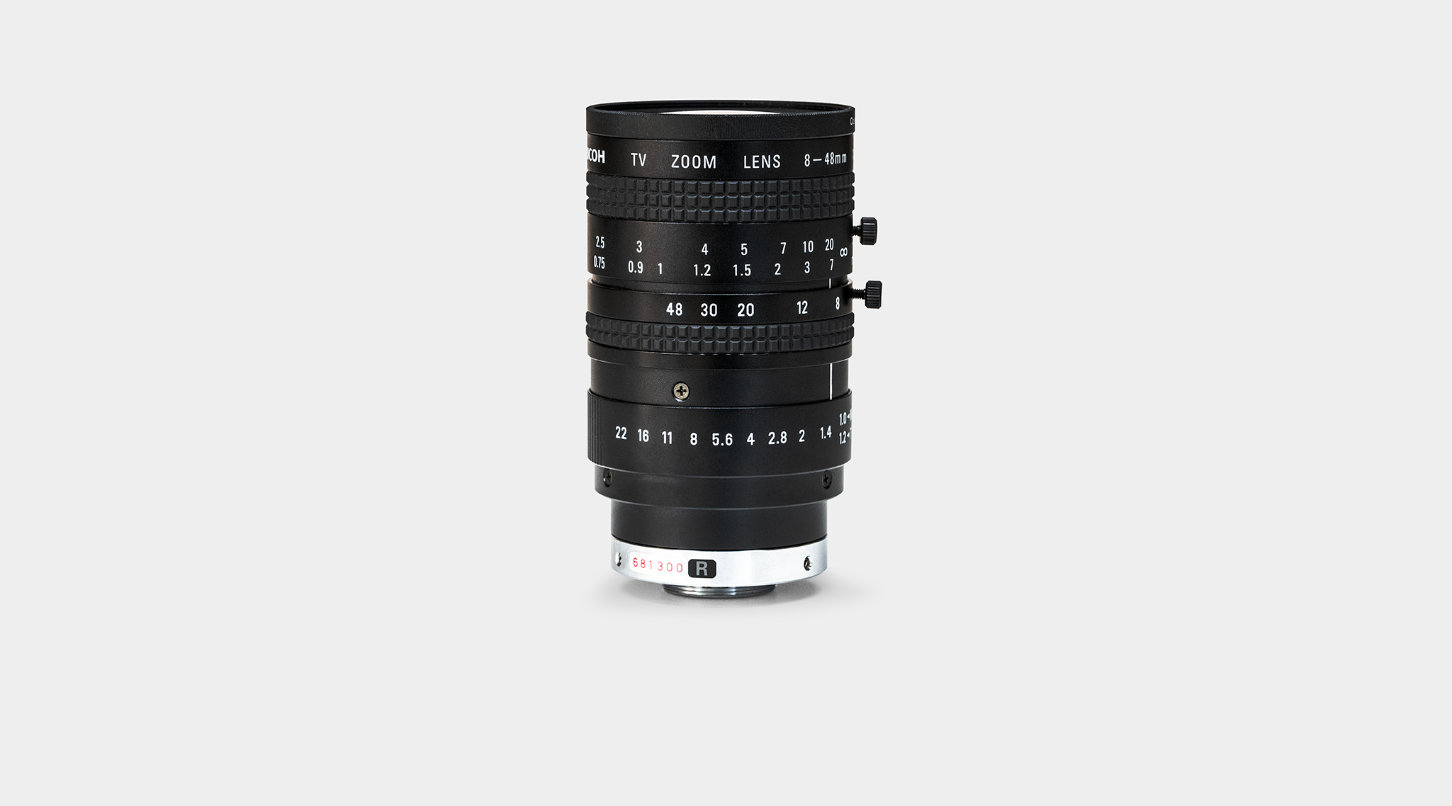
F1.8/f=12.5–75.0 mm with detector filter (RG645, 3 mm) and short-pass interference filter
51 x 90 mm
C-Mount
320 g (lens only)
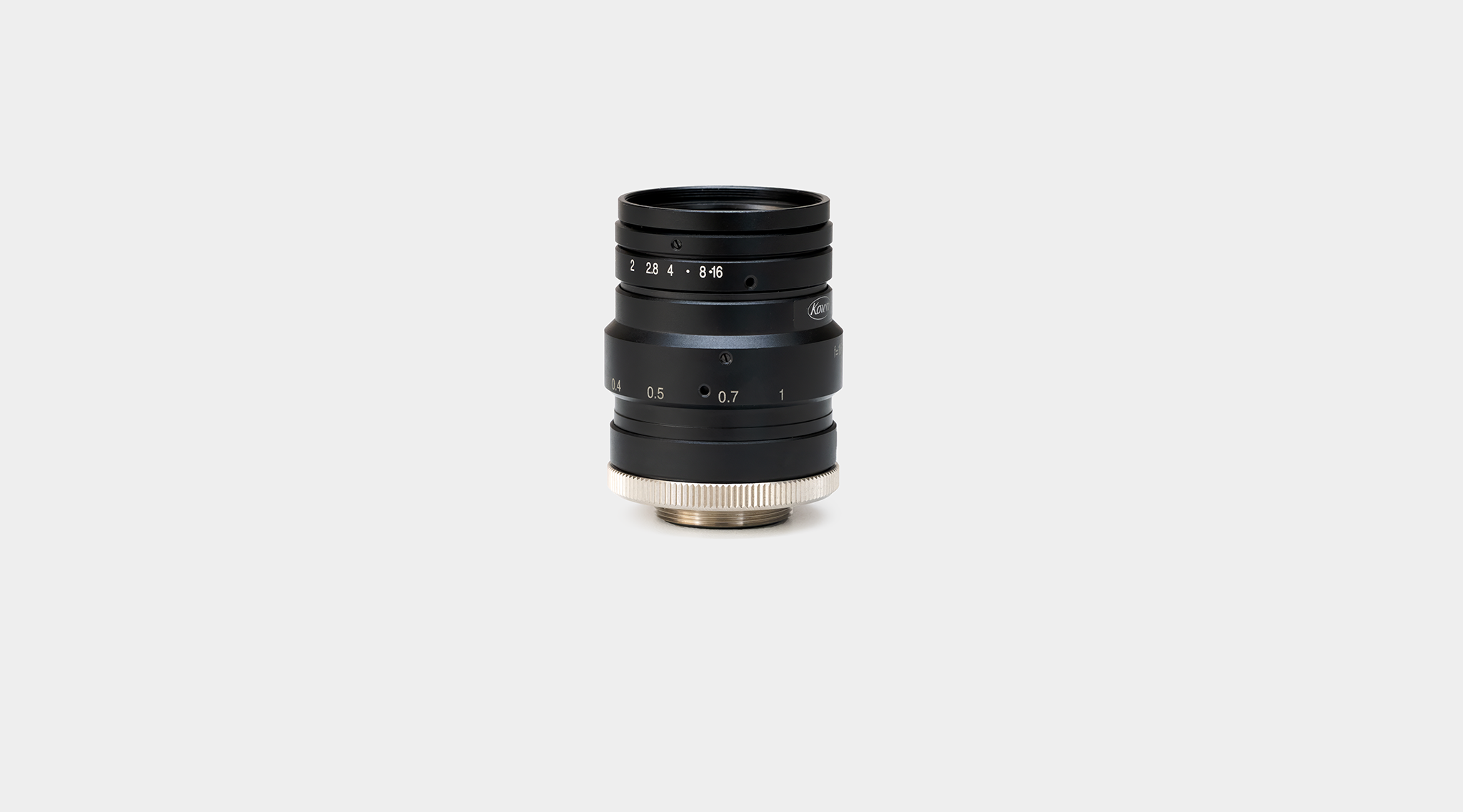
F1.4/f=25 mm with detector filter (RG645, 3 mm) and short-pass interference filter
34 x 54 mm
C-Mount
100 g (lens only)
Base plate, 40 cm x 30 cm
73.5 cm, diameter 1.5 cm
2.8 kg
Light grey plastic housing with connectors, membrane keyboard and a white illuminated LCD graphic display
12 x 7.5 x 3.5 cm
210 g (including 4 AAA 1.5 V batteries)
4 AAA-type batteries or 5 V DC from USB voltage source when connected to the computer
up to 85 % rH (avoid condensation), - 20° to + 50°C ambient temperature
PAR channel #1: 100 samples / second, PAR channel #2 and other channels: 5 samples / second (connected to computer running WinControl-3 software)
10 days or ca. 100 days automated logging with sleep mode (1 meas. / 5 min). Unlimited working time via USB connection (PC-software WinControl-3 – no sleep mode)
- Two BNC-connectors for the connection of two PAR-sensors with individual calibration factors between -50.0 and -9999.9 (memory for 10 calibration factors), range switchable in 5 steps 250 nA to 0.6 mA
- Connector for Monitoring Leaf-Clip JUNIOR-B; an adapter is available for the connection of the Leaf-Clip Holder 2030-B, and the Micro Quantum/Temp.-Sensor 2060-M
- Connector for additional digital sensors (still under development)
- USB-Connector for connection with computer (software: WinControl-3)
Flash memory used as ring buffer for 50000 lines (1 line / single measurement)
White illuminated graphic display with 5 different display modes (1: all data; 2-4: two selected sensors in big letters; 5: chart mode for channel no. 1, with maximum, minimum and average indicated), resolution: 0.1 μmol m-2 s-1
1 free USB socket. Processor, 1 GHz. RAM, 256 MB. Hard disc space, 20 MB. Screen resolution: 800 x 600 pixels. Interface, USB 1.1, 2.0 and 3.0. Operating system: Microsoft Windows 10 and 11.
Small versatile waterproof mini quantum sensor for selective PAR measurement, cosine corrected for light incident at an angle between -30 ° to +30 ° from surface normal for PPFD (photosynthetical photon flux density) measurement, with base plate for screw connection.
Black resin material
Perspex
High stability silicone photovoltaic detector with filter set for PAR correction (see “General Features” for typical response)
0.01 %/K
BNC
Base plate: 12 mm x 7 mm x 1 mm (H x W x L)
Sensor housing 5 mm x 7.5 mm x 7 mm (H x W x L)
Diffuser diameter: 3 mm
26 g
Microscopy quantum sensor for PPFD (photosynthetical photon flux density) measurement.
Black anodized aluminum housing
Resin with fluorescence dye
High stability silicon photovoltaic. Signal output typically 1.5 μA / (1000 μmol m-2 s-1)
0.01 %/K
± 5 %
0 °C … + 40 °C
55 cm
BNC
Not required
Height: 5.5 mm
Length: 76 mm
Width: 26 mm
Diffuser diameter: 0.2 mm
Stable tripod for mounting the Standard Measuring Head 3010-S, the tripod fits into the GFS-3000 transport box
54 cm – 130 cm
55 cm x 12 cm x 8 cm (L x W x H)
1050 g
This mounting set is designed for the IMAG-K9 camera and is compatible with the IMAG-MAX/L and IMAGMAX/LR LED arrays. It is made of black anodized aluminum.
The set includes a camera holder and a 15-centimeter-long, 15-millimeter-diameter metal rod for mounting the LED array on a vertical stand. Use a double socket clamp and eye protection.
200 g
Chlorophyll Fluorescence and PAM Fluorometry
Chlorophyll fluorescence is a very sensitive indicator of photosynthesis. Quantitative information on the quantum yield of photosynthetic energy conversion is obtained by PAM fluorometry and the saturation pulse method (Schreiber U (2004) Pulse-Amplitude-Modulation (PAM) Fluorometry and Saturation Pulse Method: An Overview, pp. 279-319. Kluwer Academic Publishers, Dordrecht, The Netherlands).
A wide range of photosynthetic parameters can be derived from fluorescence measurements, giving insight into the physiological state of all photosynthetically active organisms, including higher plants, mosses and ferns as well as various types of algae, phytoplankton and biofilms.
Chlorophyll Fluorescence Imaging
With the advance of highly sensitive CCD cameras and extremely powerful light-emitting diodes (LEDs), the development of IMAGING-PAM fluorometers has become possible systems that not only record images of chlorophyll fluorescence but are also fully capable of delivering all relevant chlorophyll fluorescence parameters using the saturation pulse method. This enables researchers to obtain detailed images of photosynthetic activity and to track its spatio-temporal variations with high precision. The newest addition to this technology platform, the HEXAGON-IMAGING system, is equipped with far-red (FR) LEDs that open up advanced experimental capabilities, including state-shift experiments and accurate Fo′ determinations.
All IMAGING-PAM fluorometers provide images for 17 different parameters. The fluorescence parameter Ft is monitored continuously, while Fo and Fm are determined after dark adaptation and serve as key references for fluorescence quenching analysis using the saturation pulse method. In addition to Fv/Fm, the maximum quantum yield of PS II after dark acclimation, the systems also deliver images of the effective PS II quantum yield during illumination (Y(II)), the quantum yields of regulated and non-regulated energy dissipation (Y(NPQ) and Y(NO)), as well as the apparent electron transport rate (ETR and PS).
A dedicated routine for measuring a PAR-absorptivity image is available for both the MAXI and MINI versions of the IMAGING-PAM. This “Abs.-image,” based on NIR and red-light remission, allows for direct calculation of the apparent rate of photosynthesis for each pixel—eliminating the need to rely on the commonly used universal PAR-absorptivity mean value of 0.84 (ETR). In addition, the parameter PS/50 is displayed to visualize apparent photosynthesis using the same intuitive false-color coding employed for all other photosynthesis parameters.
ImagingWin Software
General Features and Graphical User Interface
The IMAGING-PAM M-Series GigE instruments are fully controlled by the ImagingWinGigE software. When started, the ImagingWinGigE software opens with the image window that occupies most of the user surface showing the Ft value as starting parameter.
Values are represented in a false color scale ranging from black (0.0) to white (1.0) with red, orange yellow, blue and violet to purple in between. At first a standard AOI (area of interest) is already present after the start of the software. Different shapes and up to 100 AOIs can be defined. AOIs` positions can be moved by the new Edit function.


The customer can chose between 18 different parameters (Ft, Fo, Fm, F, Fm’, Fv/Fm, Y(II), Y(NPQ), Y(NO), PS/50, Abs, Red, NIR, NPQ/4, qN, qP, qL, Inh.) that could be displayed in the image window in different color modes. In this tab the alteration of the parameters can be observed in real-time during the experiment.
The kinetics window shows various parameter values for some or all AOIs of the currently chosen experiment plotted versus time. It serves for the evaluation of dynamic dark / light phenomena (Kautsky curve or Induction curve).
Some of the possible experiments are already preset in this and the following light curve window so that also the beginner finds an easy starting point for his first experiments. For advanced users it is also possible to program script files with more complex structure.
Easy light calibration using the ULM-500 Light Meter & Logger. ImagingWinGigE in communication with the ULM-500 provides an automated light calibration routine to generate a calibrated internal light list and furthermore offers to follow an external illumination.
Some new features can be provided solely for the ImagingWinGigE software, not for the ImagingWin software suitable for FireWire camera versions of the IMAGING-PAM M-Series.
Usage
Highlights
- Homogeneous illumination of measured area
- Cost efficiency due to modular design
- Remote control interface for automation
- Accessories for countless applications
Downloads
- Software: ImagingWinGigE Software
- Software: ImagingWinGigE Software (2.56zm)
- Manuals & Documentation: IMAGING-PAM M-Series Manual