MICROSCOPY-PAM
Version:
For probing of microscopic objects down to the single chloroplast

The MICROSCOPY-PAM is an extremely sensitive non-imaging chlorophyll fluorometer, which can measure smallest spots in microscopic specimen like tissue preparations or suspensions. The fluorometer consists of a modified epi-fluorescence microscope equipped with a modulated LED light source and a photomultiplier for detection of modulated chlorophyll fluorescence.
Outstanding Properties of the MICROSCOPY-PAM
Fluorescence detection by a highly sensitive red-enhanced photomultiplier tube
Single cell analysis
Version with blue or red light available
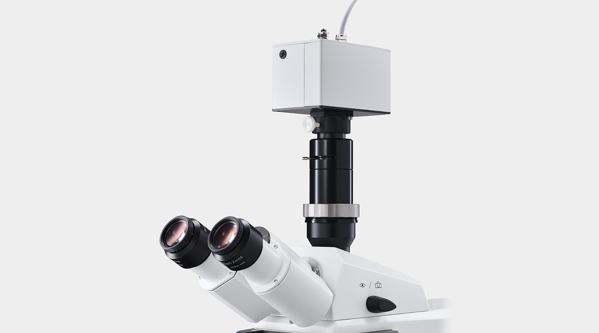


Components of the MICROSCOPY-PAM
The MICROSCOPY-PAM system uses a Zeiss AxioScope 5 epifluorescence microscope. The standard version of the microscope is equipped with a wide-aperture Zeiss Fluar objective lens with 20-fold magnification, and a blue 470 nm Zeiss LED module for fluorescence excitation. A red-orange 625 nm Zeiss LED module is available as alternative light source.
A special pulse modulation regime permits the use if the same LED module as source for measuring and actinic light as well as for saturation pulses.
Chlorophyll fluorescence is detected by a photomultiplier mounted on a special detector-ocular of the microscope. The ocular is equipped with an iris diaphragm with which the field of view can be narrowed down. A special dichroic beamsplitter filterset filters out the broadband fluorescence emission (λ > 650 nm).
Fluorescence excitation and detection are controlled by the PAM-CONTROL unit, which allows stand-alone operation of the MICROSCOPY-PAM but also functions as an interface for operation of the system by a Windows computer.
The PAM-CONTROL unit can be operated by the WinControl software versions 2 or 3. The system includes, an RS 232 cable, a USB-RS 232 adapter (when an RS-232 port is not available), a charger MINI-PAM/L, a cable to connect a chart recorder, and a transport box.
PAM-CONTROL Universal Control Unit
The PAM-CONTROL unit conducts PAM fluorescence measurements independently but it also acts as interface between fluorometer and a Windows computer running the version 2 or 3 of the WinControl software.
The PAM-CONTROL unit is part of various PAM systems, which all employ a highly sensitive photomultiplier tube for fluorescence detection: the MICROSCOPY-PAM, the MICROFIBER-PAM, and the WATER-PAM FIBER Version. In all three systems, an automatic shutdown procedure protects the photomultiplier tube against damage by high fluorescence levels or external light.
The memory of the unit can store 4000 data sets. An extensive menu provides full control of instrumental settings and a variety of measuring protocols.


Scientific Publications using Walz Devices
Source: Google Scholar.
Keywords: (Walz OR Waltz) Effeltrich.
Date: June 22, 2026.
Ʃ = 19642
Source: Google Scholar.
Keywords: (Walz OR Waltz) Effeltrich.
Date: June 22, 2026.
Ʃ = 19642
Selected Publications
Do guard cells have single or multiple defense mechanisms in response to flg22?
Physiologia Plantarum 177: e70249
Symbiont community changes confer fitness benefits for larvae in a vertically transmitting coral
Ecology and Evolution 15: e70839
Microfluidic communications in characean internodes at neutral annd alkaline external pH
Physiologia Plantarum 177: e70211
Selective permeability of Chara nodal complex for cell-to-cell passage of photometabolites exerting opposite action on chlorophyll fluorescence
Plant Physiology and Biochemistry 229: 110573
PACMan: A software package for automated single-cell chlorophyll fluorometry.
Cytometry 105: 203-213
Invasive submerged plant has a stronger inhibitory effect on epiphytic algae than native plant.
Biological Invasions 26: 1001-1014
Oscillations of chlorophyll fluorescence after plasma membrane excitation in Chara originate from nonuniform composition of signaling metabolites in the streaming cytoplasm.
Biochimica et Biophysica Acta 1865: 149019
Carbon and nitrogen uptake through photosynthesis and feeding by photosymbiotic Acantharia.
Open Research Europe
Plasma membrane-chloroplast interactions activated by the hyperpolarizing response in characean cells.
Plant Physiology and Biochemistry 201: 107836
Effects of cell excitation on photosynthetic electron flow and intercellular transport in Chara.
Protoplasma 260: 131-143
Kleptoplast distribution, photosynthetic efficiency and sequestration mechanisms in intertidal benthic foraminifera.
ISME Journal 16: 822-832
Induction changes of chlorophyll fluorescence in Chara cells related to metabolite exchange between chloroplasts and cytoplasmic flow.
Biochemistry (Moscow) Supplement Series A: Membrane and Cell Biology 15: 184-194
Effects of chloroplast-cytoplasm exchange and lateral mass transfer on slow induction of chlorophyll fluorescence in Characeae.
Physiologia Plantarum 173: 1901-1913
Cell-by-cell estimation of PAH sorption and subsequent toxicity in marine phytoplankton.
Chemosphere 259: 127487
Effects of light and daytime on the regulation of chitosan-induced stomatal responses and defence in tomato plants.
Plants 9: 59
Cell-by-cell estimation of PAH sorption and subsequent toxicity in marine phytoplankton.
Chemosphere 259: 127487
Non-photochemical quenching, a non-invasive probe for monitoring microalgal grazing: an early indicator of predation by Oxyrrhis marina and Euplotes sp.
Applied Phycology 1: 20-31
Inhibition of endosomal trafficking by brefeldin A interferes with long-distance interaction between chloroplasts and plasma membrane transporters.
Physiologia Plantarum 169: 122-134
Family-level variation in early life-cycle traits of kelp.
Journal of Phycology 55: 380-392
Cytoliths of Parietaria judaica can serve as an internal source of CO2 for photosynthetic assimilation when stomata are closed.
Journal of Experimental Botany 70: 5753-5763
Cyclosis-mediated intercellular transmission of photosynthetic metabolites in Chara revealed with chlorophyll microfluorometry.
Protoplasma 256: 815-826
Chloroplasts of symbiotic microalgae remain active during bleaching induced by thermal stress in Collodaria (Radiolaria).
bioRxiv
Mitigation of coral bleaching by antioxidants.
bioRxiv
Long-range interactions of Chara chloroplasts are sensitive to plasma-membrane H+ flows and comprise separate photo- and dark-operated pathways.
Protoplasma 255: 1621-1634
Phototrophic microbes form endolithic biofilms in ikaite tufa columns (SW Greenland).
Environmental Microbiology 19: 4754-4770
Leaf anatomy and photosynthetic efficiency of Acrostichum danaeifolium after UV radiation.
Photosynthetica 55: 401-410
Salinity effects on chloroplast PS II performance in glycophytes and halophytes.
Functional Plant Biology 43: 1003-1015
Coral disease physiology: the impact of Acroporid white syndrome on Symbiodinium
Coral Reefs 27: 373-377
Action potential in Chara cells intensifies spatial patterns of photosynthetic electron flow and non-photochemical quenching in parallel with inhibition of pH banding
Photochemical & Photobiological Sciences 7: 681-688
Effect of plasma membrane excitation on spatially distributed H+ fluxes, photosynthetic electron transport, and non-photochemical quenching in the plant cell.
In: EM Bernstein (ed) Bioelectrochemistry Research Developments, Nova Science Publishers, pp 159-188
Does the coral host protect its algal symbionts from heat and light stresses?
Proceedings of the 11th International Coral Reef Symposium, Ft. Lauderdale, Florida, 7-11 July 2008: 113-117
Compositional and fluorescence characteristics of the giant diatom Ethmodiscus along a 3000 km transect (28°N) in the central North Pacific gyre.
Deep Sea Research Part I: Oceanographic Research Papers 54: 1273-1288
The use of pulse-amplitude modulated (PAM) fluorescence-based methods to evaluate effects of herbicides in microalgal systems of different complexity
Toxicological & Environmental Chemistry 89: 665-681
Action potential in a plant cell lowers the light requirement for non-photochemical energy-dependent quenching of chlorophyll fluorescence
Biochimica et Biophysica Acta (BBA) - Bioenergetics 1767: 781-788
Guard cells in albino leaf patches do not respond to photosynthetically active radiation, but are sensitive to blue light, CO2 and abscisic acid.
Plant, Cell & Environment 29: 1595-1605
The role of phytoplankton size on photochemical recovery during the southern ocean iron experiment.
Journal of Phycology 42: 1016-1027
Fluorescence and photosynthetic competency in single eggs and embryos of Ascophyllum nodosum (Phaeophyceae).
Phycologia 45: 331-336
Photosynthetic characteristics of the phytoplankton in the Scheldt estuary: community and single-cell fluorescence measurements.
European Journal of Phycology 41: 425-434
Differential effects of plasma membrane electric excitation on H+ fluxes and photosynthesis in characean cells
Bioelectrochemistry 69: 209-215
Temporal patterns in effective quantum yield of individual zooxanthellae expelled during bleaching.
Journal of Experimental Marine Biology and Ecology 316: 17-28
Single-cell pulse amplitude modulation fluorescence measurements of the giant diatom Ethmodiscus (Bacillariophyceae)
Journal of Phycology 40: 1052-1061
Acclimation of Antarctic bottom-ice algal communities to lowered salinities during melting.
Polar Biology 27: 679-686
Effect of a single excitation stimulus on photosynthetic activity and light-dependent pH banding in Chara cells.
Journal of Membrane Biology 202: 11-19
Spatio-temporal patterns of photosystem II activity and plasma-membrane proton flows in Chara corallina cells exposed to overall and local illumination.
Planta 218: 143-151
Use of Cell-Specific PAM-Fluorometry to Characterize Host Shading in the Epiphytic Dinoflagellate Gambierdiscus toxicus.
Marine Ecology 23: 127-140
Parallel recordings of photosynthetic electron transport and K+-channel activity in single guard cells.
The Plant Journal 32: 623-630
Zooxanthellae expelled from bleached corals at 33°C are photosynthetically competent.
Marine Ecology Progress Series 220: 163–168
Measurement of light and pH dependence of single-cell photosynthesis by fluorescence microscopy.
Journal of Fluorescence 10: 269-274
New approach of monitoring changes in chlorophyll a fluorescence of single guard cells and protoplasts in response to physiological stimuli.
Plant, Cell & Environment 22: 1057-1070
MICROSCOPY-PAM
Accessories
Microscopy quantum sensor for PPFD (photosynthetical photon flux density) measurement.
Black anodized aluminum housing
Resin with fluorescence dye
High stability silicon photovoltaic. Signal output typically 1.5 μA / (1000 μmol m-2 s-1)
0.01 %/K
± 5 %
0 °C … + 40 °C
55 cm
BNC
Not required
Height: 5.5 mm
Length: 76 mm
Width: 26 mm
Diffuser diameter: 0.2 mm
WinControl-3 Software
General Features and Graphical User Interface
The MICROSCOPY-PAM can be operated by Windows computers running the WinControl-3 software. The same software operates the fluorometers DIVING-PAM-II, MICRO-PAM, MINI-PAM-II, MONITORING-PAM, and JUNIOR-PAM, PAM fluorometers operated via the PAM-CONTROL interface (MICROSCOPY-PAM, MICROFIBER-PAM and WATER-PAM FIBER-Version) as well as the Universal Light Meter ULM-500.


WinControl-2 Software
General Features and Graphical User Interface
The software WinControl-2 was developed for on-line operation via the interface PAM-CONTROL of the fluorometers MICROSCOPY-PAM, MICROFIBER-PAM, and WATER-PAM FIBER Version. WinControl-2 logs fluorescence data and automatically calculates fluorescence ratio parameters derived from saturation pulse analysis.
The WinControl-2 software runs only on Windows 7 32 bit or older Windows 32 bit operating systems.


Usage
Highlights
- Highly sensitive
- Single cell analysis
- Blue and red light versions available
Downloads
- Manuals & Documentation: JUNIOR-PAM Manual
- Manuals & Documentation: CHLORO.pdf
- Manuals & Documentation: PAM-CONTROL Manual